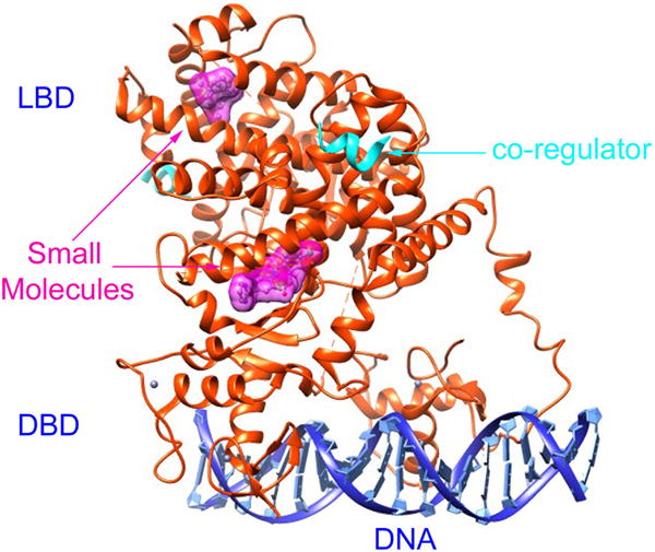
Figure 1.

An example of the TF-small molecule complex: Peroxisome proliferator-activated receptor γ (PPAR γ) complexed with DNA (PDB id: 3DZU). The heterodimer of the TF is plotted in ribbon diagram (orange). Each TF consists of a DBD and a LBD. The two DBDs bind to a sequence-specific DNA (blue). The two LBDs bind with two small molecules, represented in surface diagram (magenta), and two co-regulators, shown in ribbon diagram (cyan). The figure is created by the Chimera program.[14]
