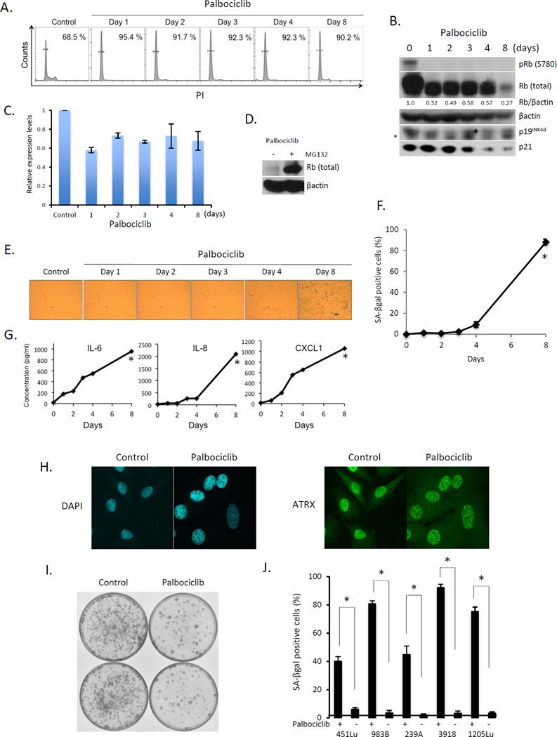
Fig. 1. Palbociclib treatment induces senescence in melanoma-derived cells.
(A) FACS analysis of 1205Lu cells treated with palbociclib. (B) Western analysis of the samples from (A). * indicates nonspecific band. (C) QPCR analysis of samples from (A). (D) Western analysis of 1205Lu cells with or without MG132 following palbociclib exposure for 8days. (E) SA-βgal staining of 1205 Lu cells following palbociclib exposure for the indicated intervals. (F) Quantification from (E) (*p < 0.001). (G) Cytokine secretion following treatment of 1205Lu cells with palbociclib (*p < 0.001). (H) DAPI staining for SAHF (left panel) and ATRX staining (right panel) in 1205Lu cells +/− palbociclib for 8 days. (I) Clonogenic assay of 1205Lu cells treated +/− palbociclib for 8 days. (J) Quantification of SA-βgal positive WM451Lu, WM983B, WM239A, WM3918, and 1205Lu cells following treatment with palbociclib for 8 days (*p < 0.001).

