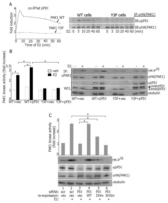
Figure 5.
βPIX facilitates E2-dependent PAK1 activation. (A) IP’d PAK1 from WT and Y3F clones treated with E2 was assessed for endogenous βPIX. (B) βPIX was overexpressed in WT and Y3F clones and PAK1 kinase activity was assessed. * indicates longer exposure with αβPIX revealing endogenous βPIX (lower double-arrow). Overexpressed βPIX is indicated by the upper double-arrow. (C) The cells were transfected with control (scr; lanes 1–2) or βPIX siRNA (lanes 3–4). In siRNA rescue experiments, βPIX DHm(L238R/L239S) (lane 5) or SH3m(W43K) (lane 6) mutants were re-expressed and PAK1 activation was assessed in the in vitro kinase.

