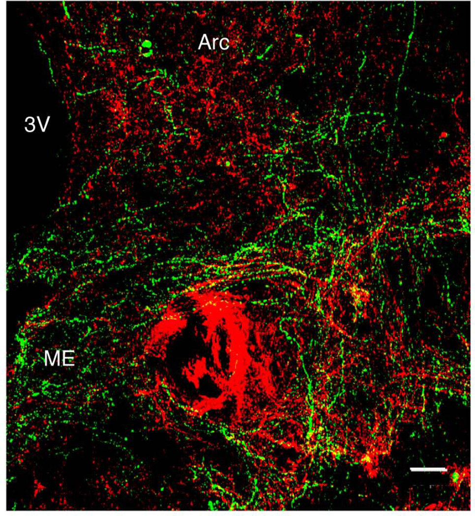
Figure 7.
Confocal projection (10×; 1 µm optical sections) illustrating the relationship between immunopositive substance P (red) and kisspeptin (green) fiber projections to the median eminence (ME) in a coronal hemi-hypothalamic section at a mid-tuberal level of a castrate adult male monkey. Substance P fibers project to the ME, and at this level clusters of substance P and kisspeptin beaded axons were observed running in close association in a near horizontal plane. Double labeled fibers were not observed. 3V, third ventricle. Arc, arcuate nucleus. Scale bar, 50µm. See also Supplementary Video to examine individual optical sections.

