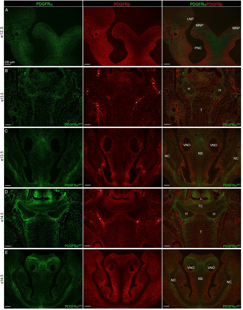
Figure 6. Pdgfrα and Pdgfrβ craniofacial expression.
(A) e12.5 (B–C) e13.5 and (D–E) e14.5. (A) PDGFRα (green) and PDGFRβ (red) protein expression in 10 µm transverse sections. (B–E) fluorescence detection of nuclear PDGFRαGFP transgene (green) and PDGFRβ protein (red) through (B, D) tongue and hyoid region and (C, E) frontonasal region. Overlap shows that the hyoid and frontonasal cartilages are devoid of Pdgfrβ expression while Pdgfrα expression is present. Asterisks (*) indicate blood vessels surrounded by Pdgfrβ expression exists. LNP=Lateral nasal process, MNP=medial nasal process, PMN= Primitive nasal capsule, H=Hyoid, T=Tongue, L= Laryngeal Aditus, TC= Thyroid Cartilage, VNO=Vomeronasal organ (Jacobson’s organ), NS=Nasal Septum, NC=Nasal Capsule.

