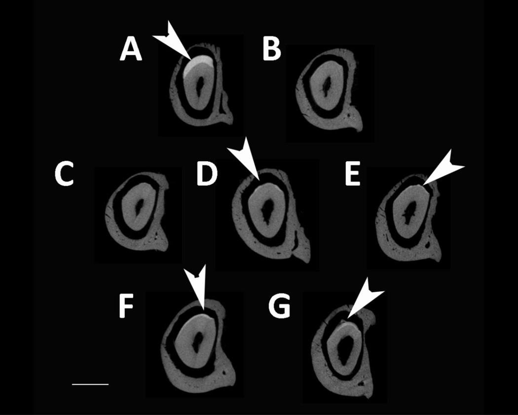
Fig. 5. MicroCT analysis of adult incisor maturation stage enamel.
MicroCT scanned sections were selected in the same location developmentally in each mouse mandible: prior to eruption in the maturation-stage of the incisor in the final section that shows the incisors completely surrounded by alveolar bone. Arrows point to mineralized enamel layers visible in incisors. Little to no enamel was visible in Amelx−/−, LRAPTg/Amelx−/−, CTRNCTg/Amelx−/− incisors (B, C, D). Thin mineralized enamel layers were visible in M180Tg/Amelx−/− (E), M180Tg/LRAPTg/Amelx−/− (F) and CTRNCTg/LRAPTg/Amelx−/− (G) incisors. Thicker mineralized enamel layers were only visible in WT incisors (A).

