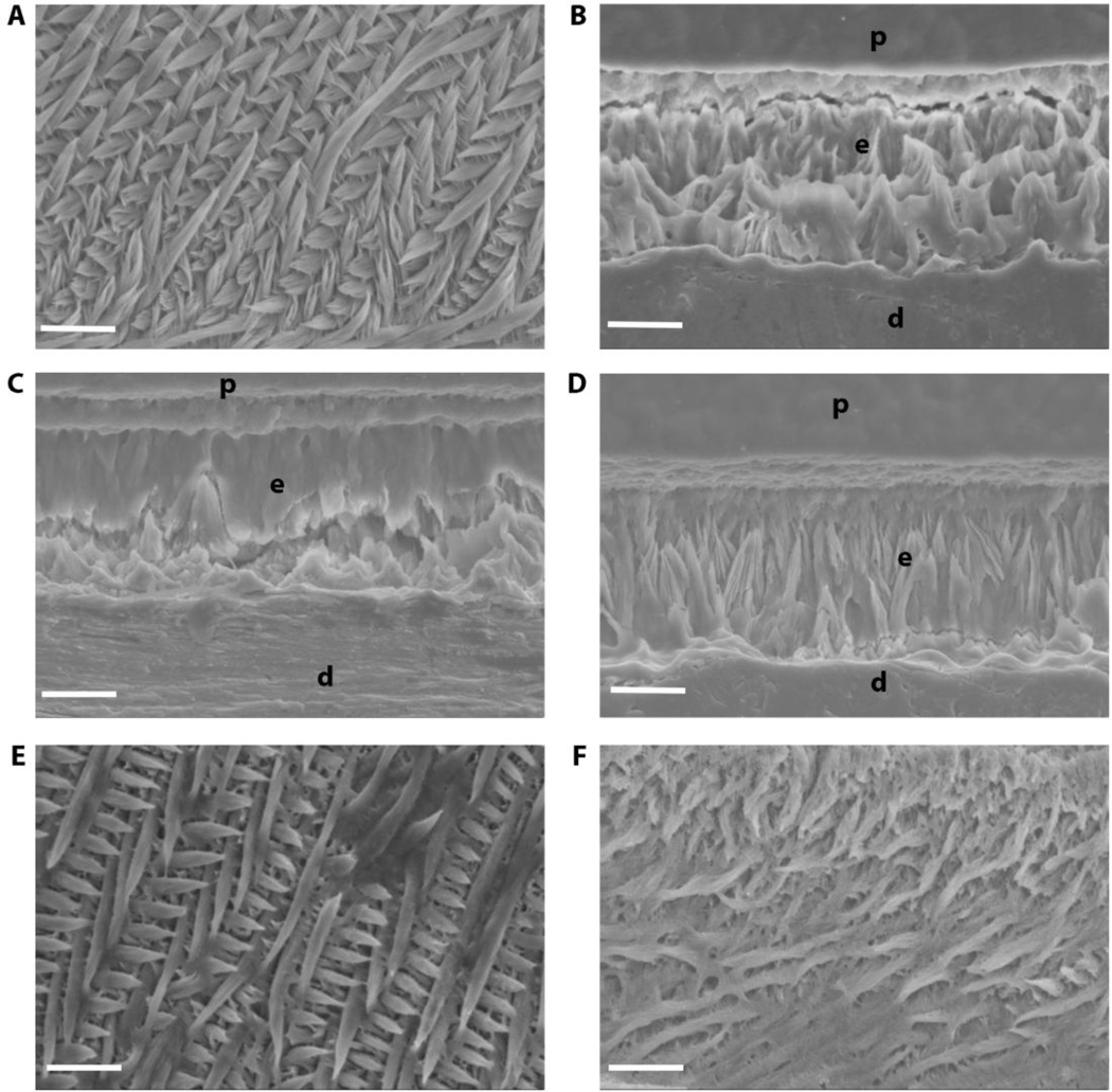
Fig. 7. Incisor enamel structure imaged by SEM.
Polished and etched longitudinal sections through mandibular incisor enamel were imaged by scanning electron microscopy in secondary mode at 2000×. While WT (A) exhibits the classic decussating prismatic organization of incisor enamel, Amelx−/− (B), LRAPTg/Amelx−/− (C), and CTRNCTg/Amelx−/− (D) incisor enamel was non-prismatic, with CTRNCTg/Amelx−/− enamel exhibiting slightly more organized mineral. CTRNCTg/LRAPTg/Amelx−/− (F) incisor enamel had noticeably more mineral organization with some decussating prisms, although not as much decussating prisms as appeared in M180Tg/LRAPTg/Amelx−/− incisor enamel (E). p = plastic embedding material, e = enamel, d = dentin

