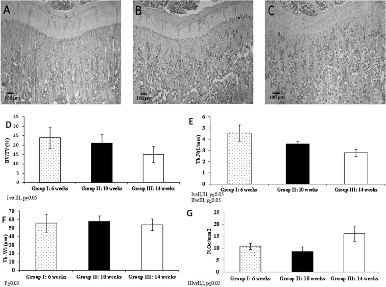
Fig. 3.
Microphotographs of tibial subchondral bone sections of different aged animals: 6-week-old animals (A); 10-week-old animals (B); 14-week-old animals (C). The images clearly show a decrease in subchondral bone volume with age. H&E, 40X. The graphs show Bone Volume (BV/TV) (%) (D), Trabecular number (Tb.N)(1/mm) (E), trabecular Width (Tb.Wi) (µm) (F) and osteoclast number (N.Oc/mm2) (G) in subchondral bone.

