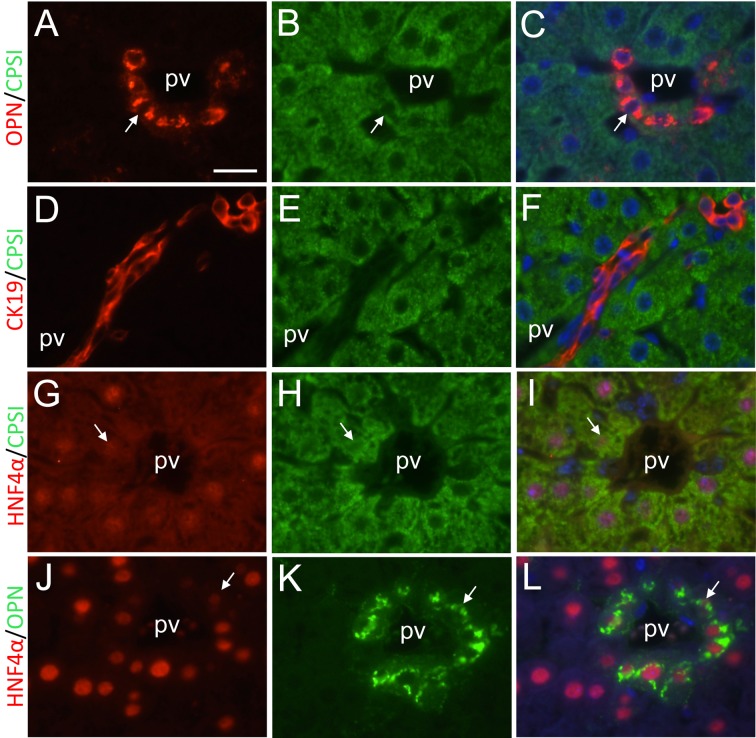
Fig. 5.
Double immunofluorescent analyses of expression of biliary and hepatocyte marker proteins. A, B, C, Osteopontin immunostaining, CPSI immunostaining and their double immunostaining at PH168, respectively. D, E, F, CK19 immunostaining, CPSI immunostaining and their double immunostaining at PH168, respectively. G, H, I, HNF4α immunostaining, CPSI immunostaining and their double immunostaining at PH144, respectively. J, K, L, HNF4α immunostaining, osteopontin immunostaining and their double immunostaining at PH168, respectively. Arrows indicate osteopontin- and CPSI-positive periportal hepatocytes (A-C), HNF4α-weakly positive and CPSI-positive periportal hepatocytes (G-I), and HNF4α-weakly positive and osteopontin-positive periportal hepatocytes (J-L). CK19-positive signals are restricted in biliary epithelial cells, and not expressed in CPSI-positive periportal hepatoctyes (D-F). pv, portal vein. Bar indicates 20 µm.

