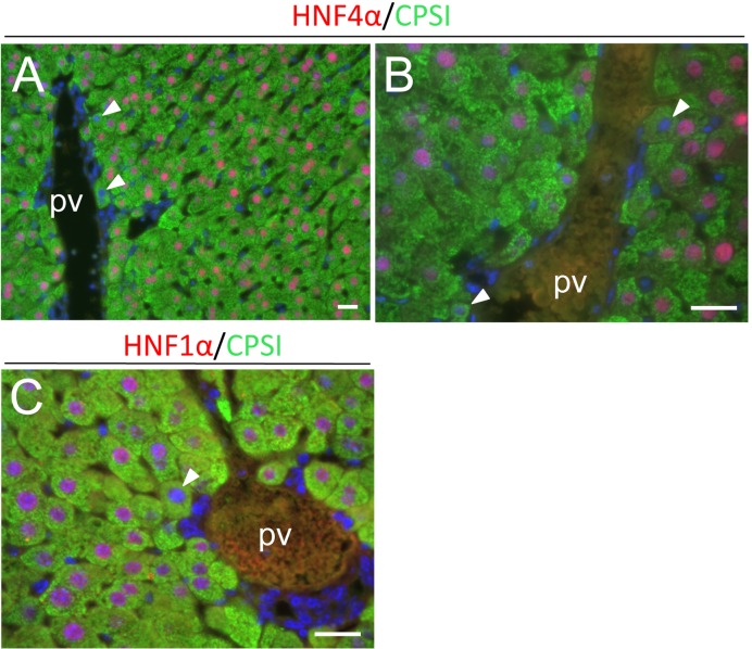
Fig. 6.
Double immunofluorescent analyses of expression of CPSI (green) and HNF4α or HNF1α (red) during liver regeneration (PH72). Nuclei were stained with DAPI (blue). Some periportal hepatocytes have HNF4α- or HNF1α-negative or very weakly positive nuclei at this time point (arrowheads)(A-C). pv, portal vein. Bars indicate 20 µm.

