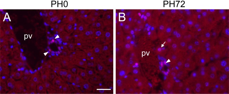
Fig. 7.
Immunohistochemical detection of nuclear localization of HES1 protein (red) in periportal hepatocytes during liver regeneration. Nuclei were stained with DAPI (blue). A, liver section at PH0. B, liver section at PH72. Nuclear localization of HES1 protein is detectable only in biliary epithelial cells (arrowheads) at PH0 (A), but a periportal hepatocyte having weakly HES1-positive nucleus (arrow) is observed in addition to biliary cells with moderately positive nuclei at PH72 (B). pv, portal vein. Bar indicates 20 µm.

