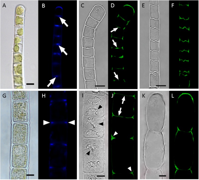
FIGURE 4.
Localization of callose in cell walls of Klebsormidium crenulatum(A–F) and Zygnema sp. Saalach (G,H). (A,B) Aniline blue staining, (C,D) staining of turgescent cell with antibody 400-2, (E,F) staining of desiccated cells with antibody 400-2, (G,H) aniline blue staining, (I,J) staining of turgescent cells with antibody 400-2, (K,L) staining of desiccated cell with antibody 400-2. Arrows: cross walls, arrowheads: cell corners. Bars 10 μm. reprinted from Herburger and Holzinger (2015).

