Figure 1.

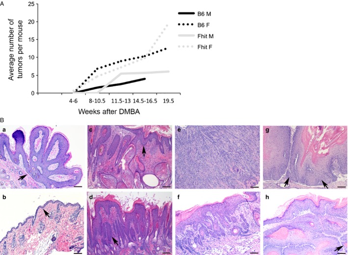
Timing of tumor induction and histopathology of DMBA/PMA‐induced skin tumors in mice with or without Zn supplementation. (A) Line graph showing the average number of tumors in wt and Fhit −/− male and female mice 4–19.5 weeks after treatment with DMBA. The data was from of a pilot study with 2–6 mice per group to estimate timing of tumor initiation in the mouse cohorts. (B) Representative photomicrographs of H&E stained skin lesions showing (a) a papilloma from a Fhit −/− female without Zn; (b) mild hyperplasia from a wt male with Zn treatment; (c) a severe hyperplasia from a Fhit −/− female with Zn treatment; (d) dysplasia from a Fhit −/− female with Zn treatment; (e) a poorly differentiated SCC with cords of highly atypical tumor cells diffusely infiltrating into the stroma and skeletal muscle from a Fhit −/− female without Zn; (f) a well‐differentiated SCC, with mild cytologic atypia from a wt female without Zn; (g) a moderately differentiated SCC, with enlarged tumor cells and prominent nucleoli, (single invasive tumor cell, left arrow; right arrow points to the tumor) from a wt male without Zn; (h) a well‐to‐moderately differentiated SCC (all portions in subcutaneous area are invasive) from a wt male with Zn treatment. Scale bar = 100 μm. Fhit, murine fragile histidine triad gene; Fhit, human or mouse protein; H&E, hematoxylin & eosin; SCC, squamous cell carcinoma; PMA, phorbol myristate acetate.
