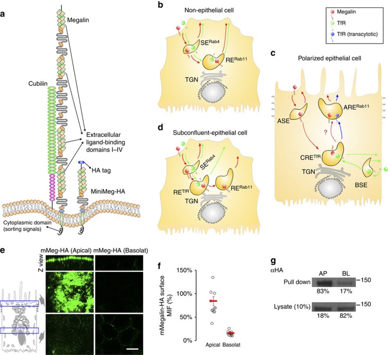
Figure 1. Model of Megalin and TfR recycling in epithelial and non-epithelial cells.
(a) Molecular representation of endogenous Megalin,Cubilin and the mMeg-HA construct. mMeg-HA contains an HA tag in the luminal domain and the entire cytoplasmic tail bearing all trafficking signals (that is, two endocytic NPxY signals and one apical sorting signal NxxY). (b) Non-epithelial cells: both Megalin and TfR are internalized into peripheral SE, where a pool of these receptors is recycled to the PM and another is transported to perinuclear RE before recycling back to the PM. (c) Polarized epithelial cells: TfR is internalized from the basolateral PM into BSE, transported to CRE and either recycled to the basolateral PM in AP-1B-positive epithelia or transcytosed to ARE in AP-1B-negative epithelia. In contrast, Megalin is internalized from the apical PM into ASE, transported to CRE, mixed with basolaterally internalized TfR, sorted to ARE and recycled to the apical PM. (d) Subconfluent epithelial cells: most Megalin is sequentially transported through three endosomal compartments (SERab4>RETfR>RERab11) before recycling back to the PM. In contrast, TfR is recycles through two endosomal compartments (SERab4>RETfR). (e) mMeg-MDCK cells polarized on Transwell filters were incubated with 488-MαHA in the apical (left) or basolateral (right) chambers (45min at 4 °C), washed (15 min at 4 °C) and fixed. Panels show Z view (top) and two confocal sections in the apical (middle) and supranuclear regions (bottom). (f) MIF quantification of the domain selective immunofluorescence described in e, where circles represent individual cells and red lines represent average and standard error. (g) Western blot analysis of mMeg-HA expression from domain selective surface biotinylation followed by streptavidin immunoprecipitation in filter-polarized mMeg-MDCK cells. Pull down (surface) and Lysate (total) fractions are shown. Scale bar, 10 μm.

