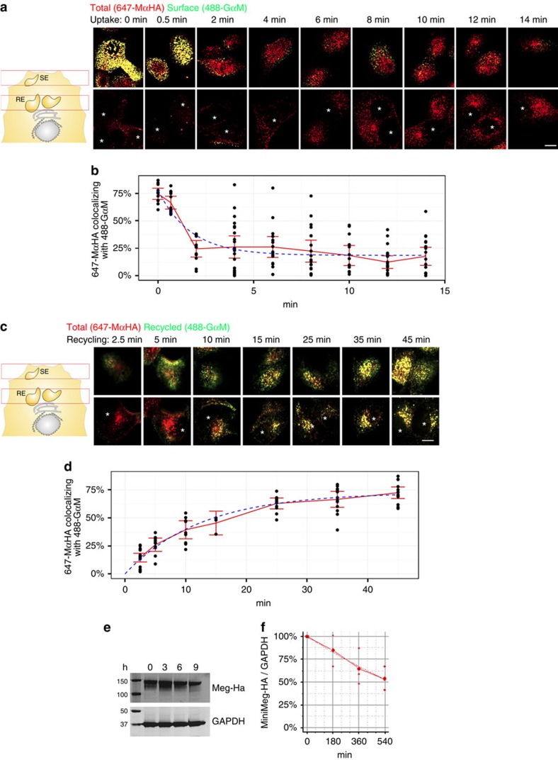
Figure 2. Megalin is endocytosed and recycled rapidly but degraded slowly in subconfluent MDCK cells.
(a) Confocal images of subconfluent mMeg-MDCK cells allowed to internalize pre-bound 647-MαHA antibody at 37 °C for the indicated times, fixed and immunostained with 488-GαM without permeabilizing. Panel show confocal sections in the upper part of the cells (top) and the perinuclear region (bottom) of the respective cells. Asterisks indicate the nuclei. (b) Co-localization quantification and fitted curve of the percentage of the 647-MαHA pixels co-localizing with the 488-GαM pixels, which informs the percentage of the total labelled mMeg-HA localized at the PM at the indicated time points. Circles represent individual cells, the red line represent average and CI95 and the blue line represents the fitted curve. (c) Confocal images of subconfluent mMeg-MDCK cells allowed to internalize 647-MαHA antibody for 45 min, washed and subsequently allowed to recycle for the indicated times in the presence of 488-GαM. (d) Co-localization quantification and fitted curve of the percentage of the 647-MαHA pixels co-localizing with the 488-GαM pixels, which informs the percentage of total mMeg-HA recycled to the PM at the indicated time points. (e) Western blot analysis of mMeg-HA and GAPDH expression in subconfluent mMeg-MDCK cells treated with cycloheximide. (f) Quantification and fitted curve of the mMeg-HA/GAPDH ratio from two experiments as the one displayed in c. Scale bar, 10 μm.

