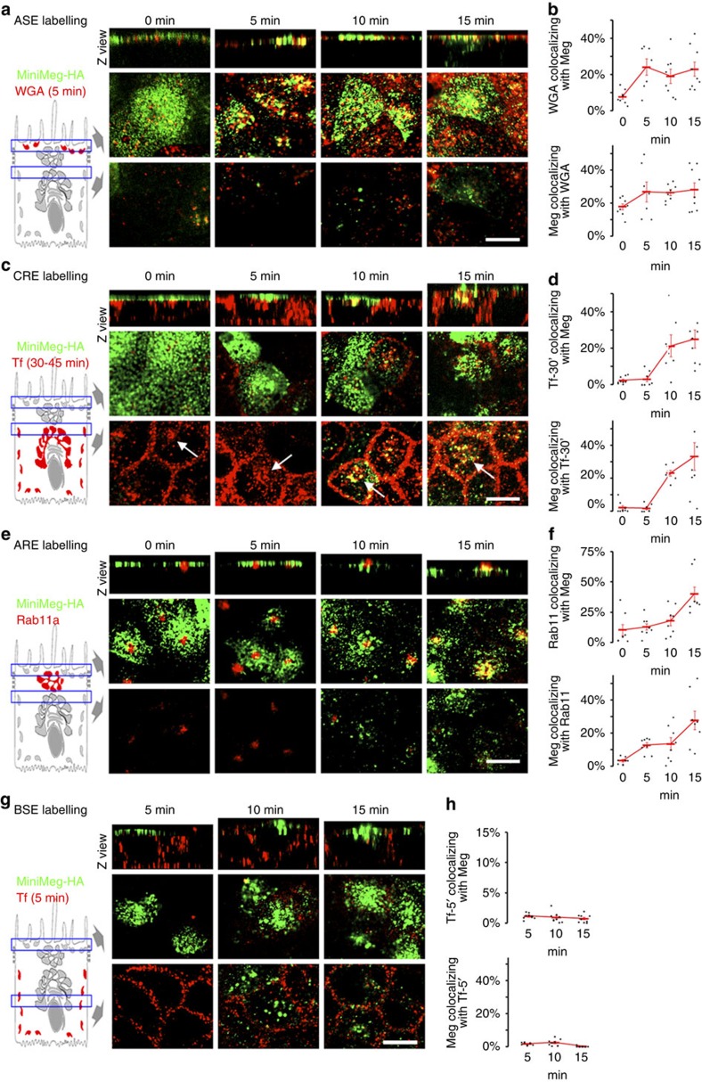
Figure 4. Megalin endosomal itinerary in polarized MDCK cells.
mMeg-MDCK cells were polarized on Transwell filters and 488-MαHA pre-bound to the apical PM was allowed to internalize for the indicated times (apical internalization assay) (a) mMeg-MDCK cells subjected to the 488-MαHA apical internalization assay were labelled for ASE with 5 min apical incubation of 594-WGA followed by stripping of the 594-WGA bound to the apical PM with N-acetyl-D-glucosamine (10 min at 4 °C, three times). Each panel displays Z view (top) and confocal sections at the level of the apical PM (middle) and supranuclear region (bottom). (b) Quantification of the percentage of the ASE marker pixels co-localizing with the 488-MαHA pixels (top) and of the percentage of the 488-MαHA pixels colocalizing with the ASE marker pixels (bottom). Circles represent individual cells, red lines represent average and s.e.. (c) mMeg-MDCK cells subjected to the 488-MαHA apical internalization assay were labelled for CRE with basolateral incubation of 594-Tf applied for 30 min. CRE appear as a subpopulation of 594-Tf-positive endosomes localized in the supranuclear region (arrows). (d) Quantification of the percentage of the CRE marker pixels co-localizing with the 488-MαHA pixels (top) and of the percentage of the 488-MαHA pixels co-localizing with the CRE marker pixels (bottom). (e) mMeg-MDCK cells subjected to the 488-MαHA apical internalization assay were labelled for ARE with anti-Rab11 antibodies. (f) Quantification of the percentage of the ARE marker pixels colocalizing with the 488-MαHA pixels (top) and of the percentage of the 488-MαHA pixels co-localizing with the ARE marker pixels (bottom). (g) mMeg-MDCK cells subjected to the 488-MαHA apical internalization assay were labelled for BSE with 5 min basolateral incubation of 594-Tf. (h) Quantification of the percentage of the BSE marker pixels co-localizing with the 488-MαHA pixels (top) and of the percentage of the 488-MαHA pixels colocalizing with the BSE marker pixels (bottom). Scale bar, 10 μm.

