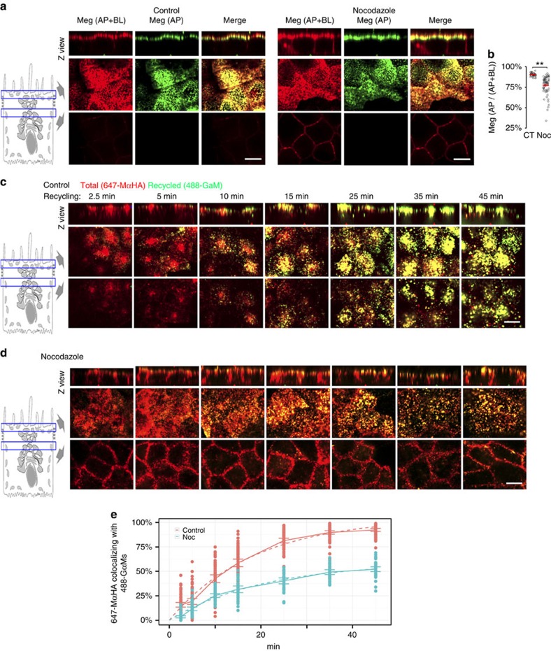
Figure 5. Microtubules mediate Megalin apical localization and recycling in polarized MDCK cells.
(a) Confocal images of control and nocodazole-treated (2 h) mMeg-MDCK cells polarized on Transwell filters and stained with 647-MαHA for the basolateral mMeg-HA and with both 647-MαHA and 488-GαM for the apical mMeg-HA. Each panel displays Z view (top) and confocal sections at the level of the apical PM (middle) and supranuclear region (bottom). (b) Co-localization quantification of the percentage of the apical mMeg-HA pixels co-localizing with the surface mMeg-HA pixels, which informs Megalin Apical/Surface ratio. Circles represent individual cells, red lines represent average and s.e., **P<0.001, two-tailed Student's t-test. (c,d) Confocal images of control (c) and nocodazole-treated (d) mMeg-MDCK cells polarized on glass-bottom chambers, allowed to internalize 647-MαHA antibody for 90 min, washed and subsequently allowed to recycle for the indicated times in the presence of 488-GαM. (e) Co-localization quantification and fitted curve of the percentage of the 647-MαHA pixels co-localizing with the 488-GαM pixels, which informs the percentage of total mMeg-HA that was recycled to the apical PM at the indicated time points. Circles represent individual cells, the continuous lines represent average and CI95 and the dashed lines represent the fitted curves. Scale bar, 10 μm.

