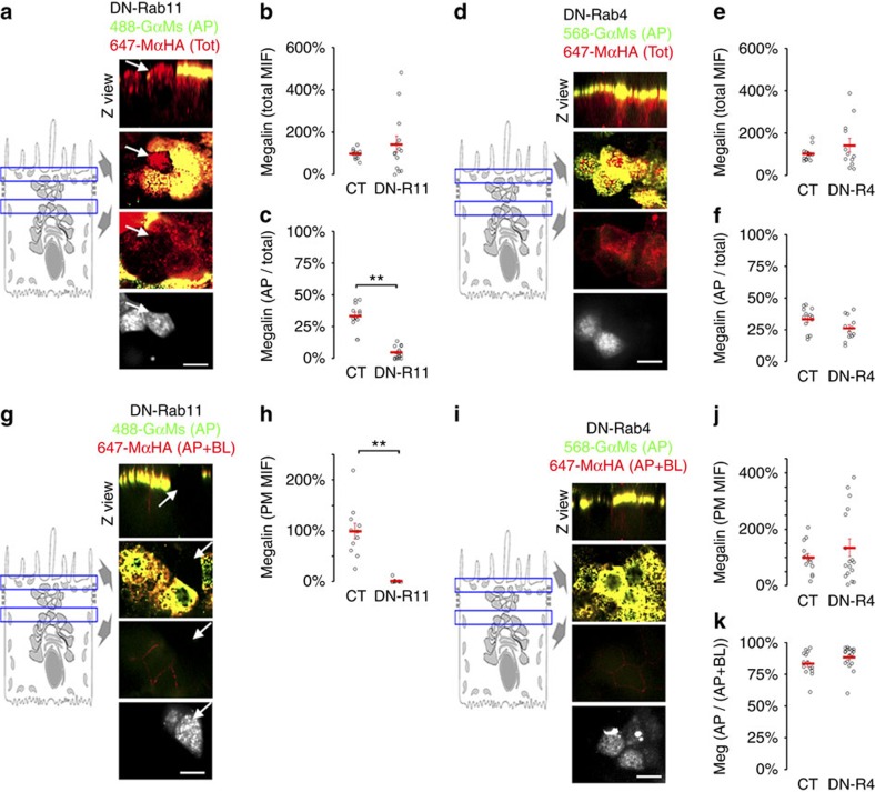
Figure 6. Rab11 mediates Megalin apical delivery in polarized MDCK cells.
(a) Confocal images of mMeg-MDCK cells polarized on Transwell filters, transiently transfected with Ch-DN-Rab11 and stained with 647-MαHA for the basolateral and intracellular mMeg-HA and with both 647-MαHA and 488-GαM for the apical mMeg-HA. Each panel displays Z view (top), confocal sections at the level of the apical PM (middle-top), supranuclear region (middle-bottom) and a supranuclear confocal section displaying the signal of Ch-DN-Rab11 (bottom). Arrows denote Ch-DN-Rab11-transfected mMeg-MDCK cells. (b) Mean intensity fluorescence (MIF) of the total mMeg-HA. (c) Percentage of the apical mMeg-HA pixels co-localizing with the total mMeg-HA pixels (apical+basolateral+intracellular), which informs Megalin apical/total ratio. **P<0.001, two-tailed Student's t-test. Circles represent individual cells, red lines represent average and s.e. (d–f) Polarized mMeg-MDCK cells were transiently transfected with GFP-DN-Rab4 and subjected to equivalent experiments to those in a,b and c. (g) Confocal images of polarized mMeg-MDCK cells, transiently transfected with Ch-DN-Rab11 and stained with 647-MαHA for the basolateral mMeg-HA and with both 647-MαHA and 488-GαM for the apical mMeg-HA. (h) Mean intensity fluorescence (MIF) of the surface mMeg-HA pool (apical+basolateral). **P<0.001, two-tailed Student's t-test. (i,j) Polarized mMeg-MDCK cells were transiently transfected with GFP-DN-Rab4 and subjected to equivalent experiments to those in g and h). (k) Quantification of the percentage of the apical mMeg-HA pixels co-localizing with the surface (apical+basolateral) mMeg-HA pixels, which informs Megalin apical/surface ratio. Scale bar, 10 μm.

