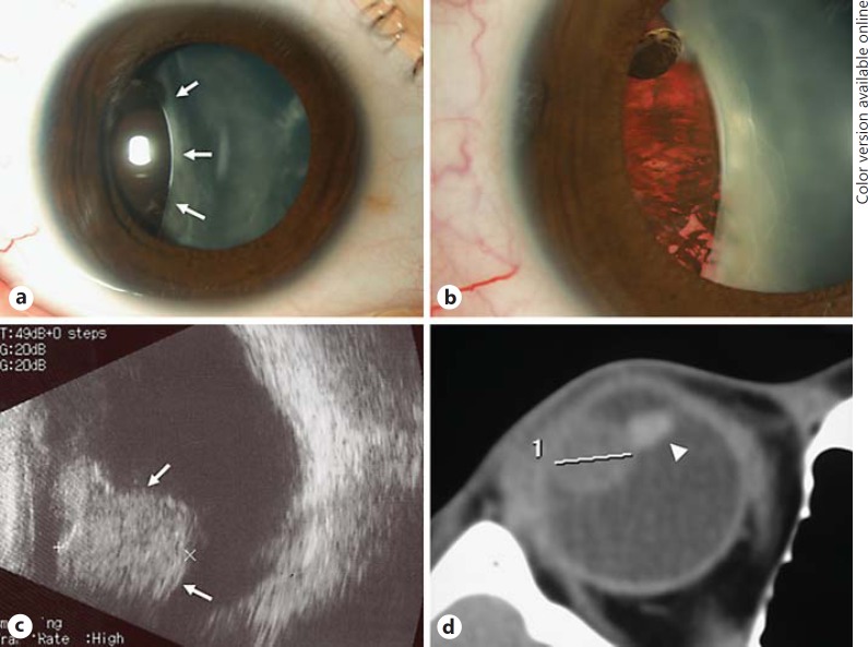
Fig. 1.
Clinical features of the tumor. a Slit-lamp photograph showing a brown tumor (arrows) behind the iris and cataract. b The temporal episcleral vessels were slightly dilated. Note the presence of a cataract with indentation of the lens caused by the tumor. c Ultrasonography demonstrated an oval and solid mass (arrows) at the ciliary body. d Axial computed tomography showed an oval tumor 11 mm in diameter at the ciliary body. Note the deformed and tilted lens caused by the tumor (arrowhead).

