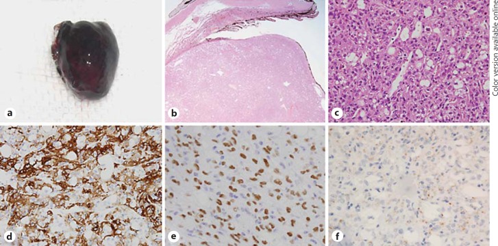
Fig. 2.
Histopathology of the excised tumor. a Gross photograph of the resected tumor. b The tumor arose from the pars plana of the ciliary body (hematoxylin-eosin, original magnification ×100). c Note the proliferated polygonal epithelioid cells with eosinophilic cytoplasm and vacuolated cells (hematoxylin-eosin, ×400). d Immunohistochemical staining showed a strong positive reaction for SMA (×400). e TFE-3 was diffusely positive in individual cells (×400). f Adipophilin was positive around vacuolated cells (×400).

