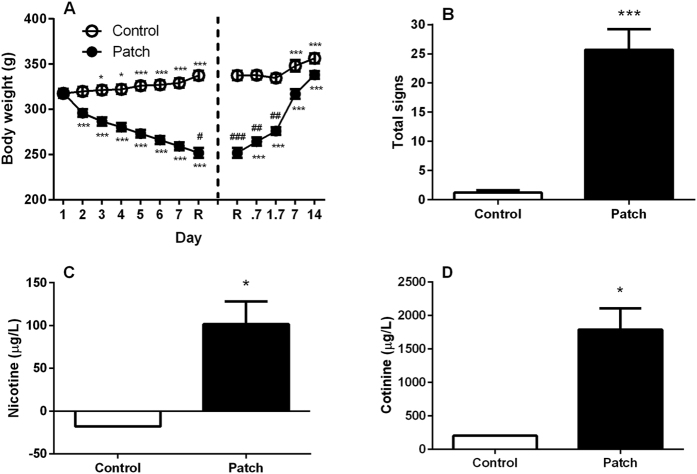
Figure 2. Dependence state upon nicotine following 7.3 days of chronic exposure to transdermal nicotine patches (5.2 mg/rat/day).
Dependence was verified (A) by measuring body weight loss and gain across the intoxication period and upon nicotine discontinuation, respectively. Data are the mean (±SEM) grams (g) of body weight during intoxication from day 1 to the time of removal (R) and at 16 hours (0.7 days), 40 hours (1.7 days) 7 and 14 days following R. *p < 0.05, ***p < 0.001 difference from day 1 or R in both nicotine exposed (patch, N = 9) and non-exposed (control, N = 9). #p < 0.05, ##p < 0.01, ###p < 0.001 difference from control. Dependence was also verified (B) by observing spontaneous withdrawal signs 16h after patch removal. Rats previously exposed to nicotine showed increased occurrence of overall withdrawal signs compared to non-exposed controls. Overall withdrawal signs were obtained for each animal by accumulating different categories of somatic withdrawal manifestations (cheek tremors, teeth chattering, head/body shakes, gasps and writhes). ***p < 0.001 difference from control. Finally, to control for effective nicotine release by patches (C) blood nicotine and (D) cotinine levels in the blood were verified two hours following the last change of patches. Nicotine exposed rats showed elevated blood nicotine and cotinine concentrations expressed as mean (±SEM) micrograms per liter (μg/L) compared to non-exposed rats. The signal detected in the control group for both nicotine and cotinine were not significantly lower or over the blank standard, respectively. *p < 0.05 difference from control. For detailed statistics, see “Results”.

