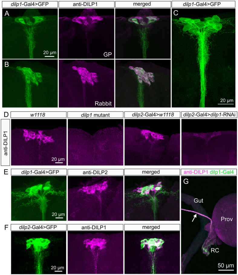
Figure 1. DILP1 is expressed in IPCs in pars intercerebralis of the dorsal brain of newly-eclosed flies.
(A,B) Antisera to DILP1 produced in guinea pig (GP; A) and rabbit (B) label neurons identified by dilp1-Gal4-driven GFP. Although labeling intensity appears somewhat variable when the two markers are used simultaneously, it is clear that all IPCs coexpress the markers. (C) Higher resolution image of the 14 IPCs expressing dilp1-GFP. (D) DILP1 immunolabeling is not detected in the brain of dilp1-mutant flies. Also after dilp2-Gal4-driven dilp1-RNAi the DILP1 immunolabeling is strongly reduced. (E) To confirm that DILP1/dilp1 is co-expressed in DILP2 producing IPCs we applied anti-DILP2 to brains with dilp1-Gal4-driven GFP and reveal colocalization of markers. (F) Also the reverse experiment with dilp2-Gal4 >GFP and anti-DILP1 showed coexpression. (G) Details of axon terminations of neurons coexpressing DILP1-immunolabeling and dilp1-Gal4-GFP expression in the foregut structures proventriculus (Prov) and retrocerebral complex (RC; corpora cardiaca and corpora allata).

