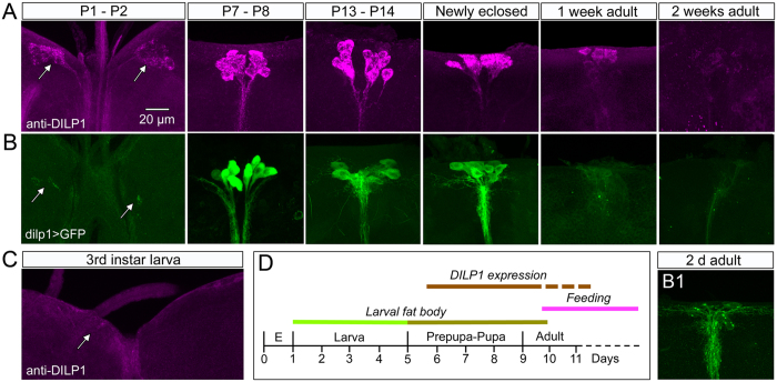
Figure 2. Temporal expression profile of DILP1 immunolabeling and dilp1-Gal4 expression in the brain.
(A) In Canton S flies DILP1 immunolabeling appears first in early pupal stages (P1-P2), remains strong in newly eclosed flies and starts fading in 1-week-old flies. (B) Dilp1-Gal4-driven GFP follows the same expression pattern until flies are newly eclosed. B1 At about 2–3 days of adult life GFP intensity starts fading. (C) In the larval brain no DILP1 or dilp1 expression could be detected. (D) Time course of DILP1 expression in relation to Drosophila development, presence of larval fat body and onset of feeding. We did not investigate DILP1 expression during embryonic (E) development.

