Figure 4.
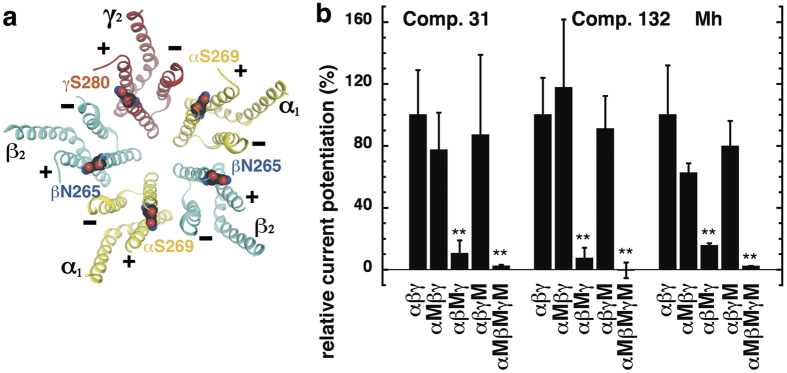
(a) Model structure of the GABAA receptor transmembrane domain. The major isoform of the GABAA receptor is composed of two α1, two β2, and one γ2 subunits. The model structure depicts the crystalized homomeric β3 GABAA receptor (PDB structure 4COF)38. In this figure, some of the β3 subunits were renamed α1 (yellow), β2 (blue) and γ2 (red); structures are shown in ribbon representation. The mutated residues α1S269, β2N265, and γ2S280 are located at the interfaces between subunits. (b) Potentiation of the GABA response by compound 31 (3 μM), compound 132 (3 μM), and 4-O-methylhonokiol (1 μM, abbreviated Mh) in wild-type α1β2γ2, single mutant (α1M, β2M, γ2M), and triple mutant receptors expressed in Xenopus oocytes. The bars indicate mean ± SD, n = 3.

