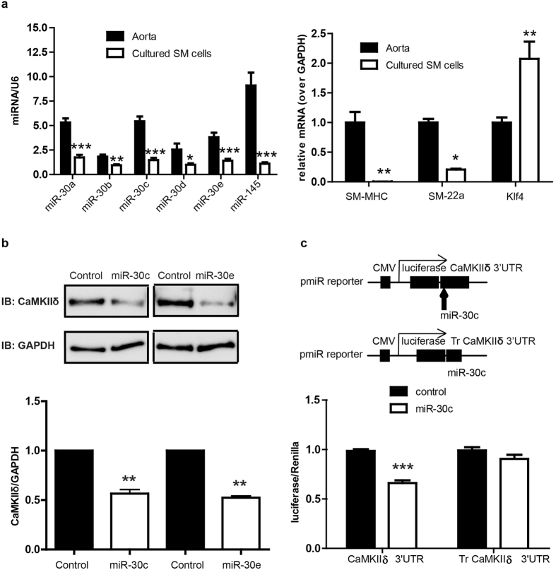
Figure 2. MiR-30 inhibits CaMKIIδ expression by targeting CaMKIIδ 3′UTR in primarily cultured smooth muscle cells.
Rat aorta medial layers were enzymatically dispersed and primarily cultured for 3–7 passages. (a) The miRNA and mRNA levels of miR-30a, miR-30b, miR-30c, miR-30d, miR-30e, SM-MHC, SM-22α and Klf4 were measured by qPCR in aorta and cultured smooth muscle cells. Data were normalized over U6 (miRNA) or GAPDH (mRNA) (n = 3 aorta and 5 cultured SM cells). (b) miR-30c mimic or miR-30e (Invitrogen) (0.2pmol) was eletroporated into primarily cultured SM cells (1 million) and the expression of CaMKIIδ and GAPDH was analyzed by Western blot 3 days post-electroporation. (c) Full length of CaMKIIδ 3′UTR or truncated CaMKIIδ 3′UTR was introduced into pmiR reporter vector (Invitrogen). Full length CaMKIIδ 3′UTR reporter or truncated CaMKIIδ 3′UTR reporter as well as miR-30c and renilla were transfected into HEK293 cells and luciferase activity was measured 3 days after transfection. Values are shown as mean +/- S.E. M., n ≥ 4 and analyzed by two-way ANOVA or t-test. *p < 0.05 **p < 0.01 and *** p < 0.001.

