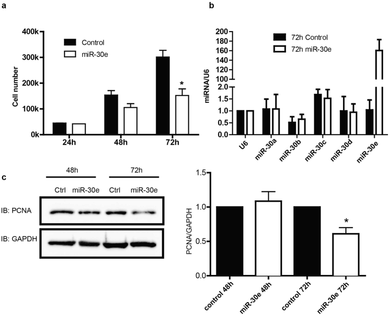
Figure 3. MiR-30 inhibits vascular smooth muscle cell proliferation.
miR-30e mimic (0.2 pmol) was eletroporated into cultured vascular smooth muscle cells (50,000). (a) Cell numbers were counted at 24 h, 48 h and 72 h post eletroporation to monitor cell growth and proliferation. (b) The level of miR-30 family members was tested using qPCR 72 h after eletroporation. (c) Cell lysates were immnuoblotting for PCNA and GAPDH and quantified data were normalized over GAPDH. Values shown are mean±S.E.M., n = 3 pairs and *p < 0.05 ***p < 0.001 analyzed by two-way ANOVA.

