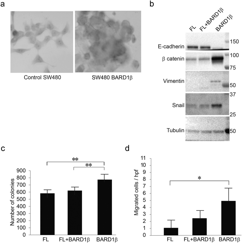
Figure 5. Expression of BARD1β is associated with a more invasive phenotype.
(a) Control (left) and BARD1β expressing (right) SW480 cells were stained with hemotoxylin, and inspected under a light microscope (400x magnification). (b) Stable SW480 cells infected with either FL BARD1, BARD1β or the combination (FL + BARD1β) were harvested, separated, and immunoblotted for EMT proteins: E-cadherin, β-catenin, vimentin, or snail. Tubulin was used as a loading control. (c) Colony formation assay of stable SW480 cells: 100 cells were seeded into six-well plates and incubated for two weeks. Colonies were fixed and stained with crystal violet (*p < 0.05 and **p < 0.01). (d) The relative number of migrated stable SW480 cells per high power field (hpf) (*p < 0.05 and **p < 0.01).

