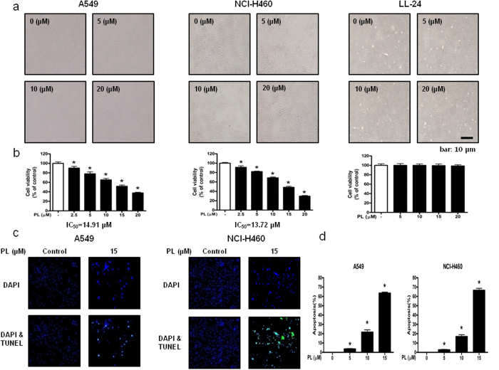
Figure 1. Effect of PL on the growth of NSCLC cells and lung epithelial cells, and effect of PL on apoptotic cell death in NSCLC cells.
Concentration-dependent inhibitory effect of PL on cancer cell growth was found in A549 and NCI-H460 NSCLC cells but not in LL-24 cells. (a) Morphological changes of A549 and NCI-H460 NSCLC cells and LL-24 lung epithelial cells were observed under phase contrast microscope. (b) Relative cell survival rate was determined by MTT assay. Data was expressed as the mean ± S.D. of three experiments. *p < 0.05 indicates significant difference from control group. (c) Apoptotic cell death of A549 and NCI-H460. NSCLC cells were treated with PL (0–20 μM) for 24 h, and then labeled with DAPI and TUNEL solution. Total number of cells in a given area was determined by using DAPI nuclear staining (fluorescent microscope). A green color in the fixed cells marks TUNEL-labeled cells. (d) Apoptotic index was determined as the DAPI-stained TUNEL-positive cell number/total DAPI-stained cell number x 100%. Data was expressed as the mean ± S.D. of three experiments. *p < 0.05 indicates significant difference from control group.

