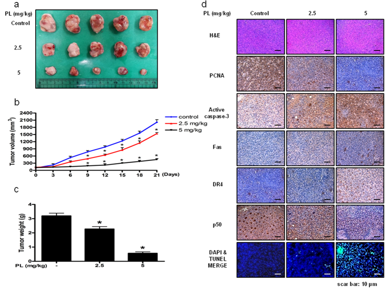
Figure 7. Anti-tumor activity of PL in lung cancer xenograft mice model.
(a–c) Growth inhibition of subcutaneously transplanted A549 xenografts mice treated with PL (2.5 mg/kg and 5 mg/kg twice a week) for 3 weeks. Xenograft mice (n = 10) were administrated intraperitoneally with 0.01% DMSO or PL (2.5 mg/kg and 5 mg/kg). Tumor burden was measured once per week using a caliper, and calculated volume length (mm) × width (mm) × height (mm)/2. Tumor weight and volume are presented as means ± S.D. (d) Immunohistochemistry was used to determine expression levels of H&E, PCNA, active caspase-3, NF-κB p50 subunit in nude mice xenograft tissues by the different treatments as described in the materials and methods. We also performed DAPI&TUNEL assay to assess the apoptosis rate in the nude mice xenograft tissues. Total number of cells in a given area was determined by using DAPI nuclear staining (fluorescent microscope). A green color in the fixed cells marks TUNEL-labeled cells.

