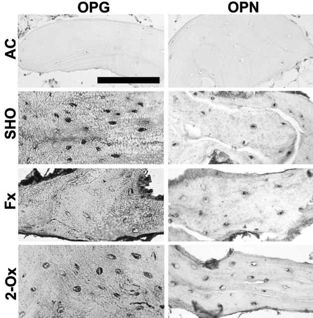
Fig. 1.

Representative pictures of the immunohistochemical analysis of the expression of osteopontin (OPN) and osteoprotegerin (OPG) carried out on formaldehyde-fixed sections from femoral trabeculae of pigs from the Fx group, the fundectomized group; the 2-Ox group, supplemented with 2-Ox after fundectomy; and the SHO group, the sham-operated group. AC, antibody control. Scale bar for all panels: 200 µm.
