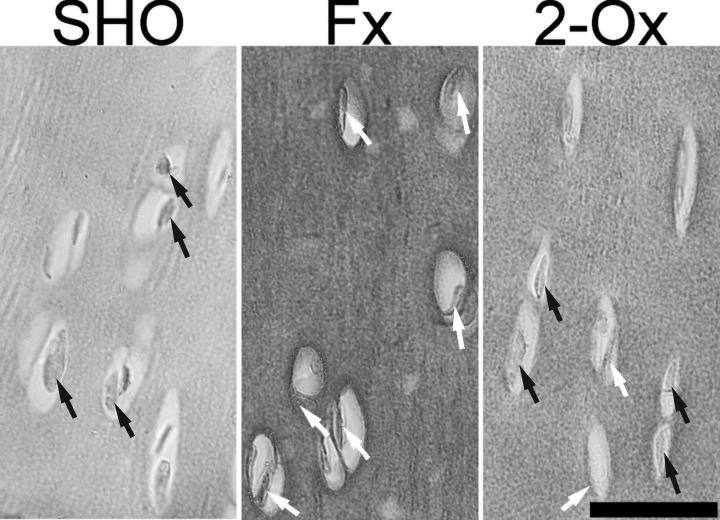
Fig. 2.
Representative pictures of the immunohistochemical analysis of the expression of osteocalcin (OCN) carried out on formaldehyde-fixed sections from femoral articular cartilage of pigs from the Fx group, the fundectomized group; the 2-Ox group, supplemented with 2-Ox after fundectomy; and the SHO group, the sham-operated group. In normal chondrocyte from the SHO group, the nucleus is stained blue (the negative OCN signal) and indicated by black arrows. The positive OCN signal is indicated in cells from the Fx group by white arrows. Scale bar: 200 µm.

