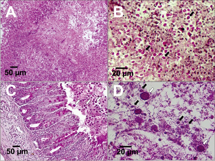
Fig. 6.
Thin sections of liver abscesses from CBA mice infected with Tritrichomonas foetus (IMC cl2) 7 days after inoculation (periodic acid–Schiff (PAS) staining) (A, B). Thin sections of the cecum of CBA mice co-infected with T. foetus (IMC cl2) and Entamoeba histolytica (HM-1:IMSS cl6). Data obtained at 5 days after inoculation via orogastric intubation are shown (PAS staining) (C, D). Black arrows: T. foetus (IMC cl2), white arrows: E. histolytica (HM-1:IMSS cl6).

