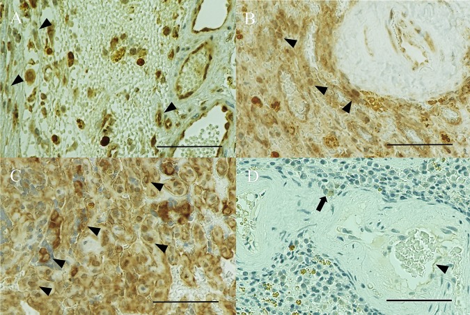
Fig. 2.
VEGFR-2 immunohistochemical staining (× 400). (A) Hemangiosarcoma (HSA) showing weak (1+) cytoplasmic and nuclear expression (arrowhead) in Case 5. (B) HSA showing strong (2+) cytoplasmic and nuclear expression (arrowheads) in Case 4. (C) HSA showing strong (3+) cytoplasmic and nuclear expression (arrowheads) in Case 1. (D) A normal spleen showing no expression (control), which may reflect the presence of VEGFR-2-expressing endothelial cells (arrowhead) and macrophages (arrow). The nuclei of lymphocytes show dense blue stain in Case 12. Bar, 60 µm.

