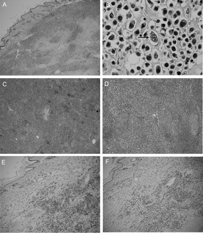
Fig. 4.
Post mortem histopathological examination of the subcutaneous mass (A, B), liver (C) and spleen (D). Tumor cells infiltrated all three tissues. Cells (B) were composed mainly of proplasmacytes with a small number of mature plasma cells (short arrow) and plasmablasts (long arrow). Hematoxylin and eosin staining. (A, C and D) × 10 objective and (B) × 100 objective. The subcutaneous mass is positive for CD79a (E) and MUM1 (F). Hematoxylin counterstain. (E, F) × 10 objective.

