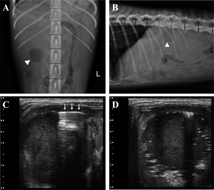
Fig. 1.
Ventrodorsal view (A) and right lateral view (B) of abdominal radiographs showing gas opacity (arrowhead) superimposed on enlarged right kidney. Small intestine in cranial abdomen is displaced ventrally to kidney with microhepatica (B). A reverberation artifact (C, arrows) and bright speckles with distal acoustic shadowing (D) existed internally interfacing the diverticuli.

