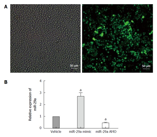
Figure 5.

Lentiviral transfection and miRNA expression after transfection. A: Cells were infected with 50 MOI of lentivirus, and imaged 72 h post-transfection. Comparison of bright field filter view to FITC filter view (GFP-expression cells) for the same fields of cells showed about 90% infection efficiency by 72 h; B: Quantitative real-time PCR analysis of miR-29a expression in the AR42J cells after transfection. Date are shown as a ratio of mi-29a mimic and AMO groups to vehicle groups using the 2-ΔΔct. Data are representative of three independent experiments. aP < 0.05 vs vehicle group.
