
Figure 6.
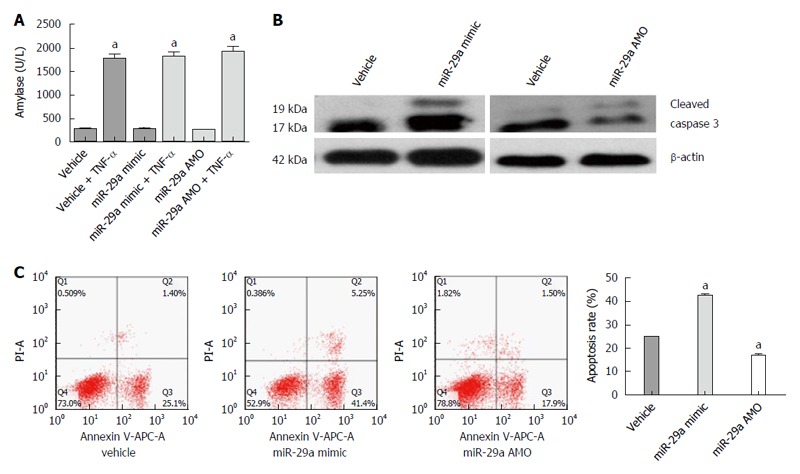
miR-29a promotes the apoptosis of the AR42J cells. A: The amylase analysis in the supernatant increased obviously; B: Western blot analysis of activated caspase 3 in AR42J cells; C: The apoptosis rate of AR42J cells was determined by FACS analysis. Data are representative of mean ± SD from three independent experiments performed in triplicate. aP < 0.05 vs control or vehicle group.