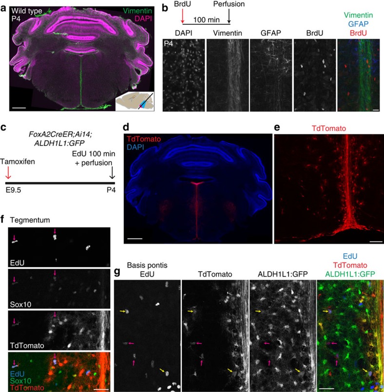
Figure 6. Proliferative progenitors in postnatal pons deriving from midline domain.
(a) Wild-type mouse pons sectioned at oblique angle and stained for vimentin (green) and DAPI (magenta). Note the intense vimentin staining along the midline. Scale bar=500 μm. (b) Pons tissue from a mouse perfused 100 min after a single dose of BrdU (50 mg kg−1) was co-stained for Vimentin, GFAP and BrdU, revealing proliferating cells among fibres of the midline. Scale bar, 20 μm. (c) Schematic of fate mapping of midline domain. (d) TdTomato labelling by FoxA2CreER;Ai14 in caudal pons, extending from fourth ventricle along midline, and including nearby cells. The midline and nearby cells were clearly labelled, along with other pontine structures known to derive from FoxA2+ progenitors (ref. 67): the motor nuclei of cranial nerves V, VI and VII, and raphe nuclei. Scale bar, 500 μm. (e) TdTomato expression in fibres along midline and in nearby parenchyma of basis pontis. Scale bar, 100 μm. (f) Co-labelling of TdTomato, EdU, and Sox10 reveals P4 proliferative oligodendroglia in tegmentum labelled by FoxA2CreER recombination at E9.5. Magenta arrows indicate Sox10+EdU+TdT+ cells. Scale bar, 50 μm. (g) Co-labelling of TdTomato, EdU, and ALDH1L1:GFP reveals that some P4 proliferative astrocytes are labelled by FoxA2CreER recombination at E9.5. Yellow arrows indicate ALDH1L1:GFP+EdU+TdT+ cells; magenta arrows indicate GFP−EdU+TdT+ cells. Scale bar, 50 μm. (d–g) In a section of bilateral width 4 mm, we observed no TdT+ EdU+ cells further than 500 μm from the midline; the only TdT+ cell bodies beyond 1 mm from midline were trigeminal motor neurons. These results show that while the midline domain produces postnatally proliferative astroglia and oligodendroglia, these progeny are regionally restricted.

