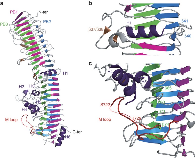
Figure 1. X-ray structure of the HxuA molecule.
(a) Cartoon representation of HxuA, a right-handed β-helix. Parallel β-sheet PB1 is coloured in magenta, PB2 in blue, PB3 in green and the extra-helix strand motifs in brown. The α-helix elements H1–H6 are shown in purple. (b) Enlarged view of α-helix H1, which splits PB1 into two parts. Together with the extra motifs β37/β38 and the swap of β40 and β41, this element induces a twist in the β-helix, highlighting the separation between the secretion domain and the functional domain. (c) Enlarged view of loop 706–731, in red, referred to as the M loop, which undergoes an important conformational change during complex formation.

