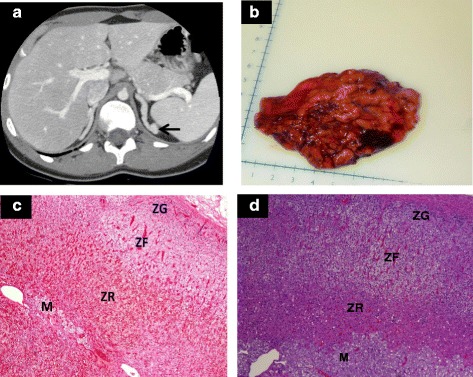
Fig. 1.

a Adrenal computerized tomography scan shows bilateral adrenal hyperplasia with a left adrenal gland nodule (arrow). b Hypertrophied right adrenal gland (weight 41.8 grams, normal adrenal weight 4 – 6 grams). c Histopathology of the classic simple virilizing CAH adrenal shows preservation of the adrenal cortex zonation (ZG, zona glomerulosa; ZF, zona fasciculata; ZR, zona reticularis) and the inner medulla (M) (d) Histology of a healthy unaffected adrenal gland shows the outer cortex with characteristic zones (Hematoxylin and eosin stain, magnification 4X)
