
Figure 5.
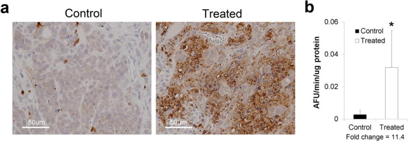
Caspase-3 activity verification in tumors from mice imaged by microPET. a Representative tumor sections were stained immunohistochemically for cleaved caspase-3 in N = 2 mice (N = 4 tumors) per treatment group. b Tumor caspase-3 enzyme activity was determined in N = 4 mice (N = 8 tumors) per treatment group. Fold-change = mean caspase-3 activity in tumors from M413-treated mice divided by mean activity of tumors from PBS-treated mice.