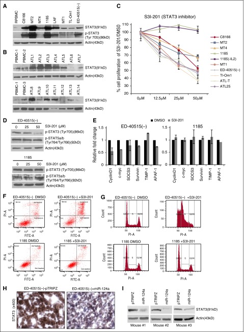
Figure 3.
HTLV-I/ATL cells over-express STAT3, which is required for cell survival. (A-B) Increased expression of STAT3 and p-STAT3 (Tyr705) in HTLV-I and ATL cell lines and increased STAT3 expression in primary ATL patient samples (n = 14). Resting noninfected PBMCs (R.PBMCs, n = 3) served as controls. Actin served as a loading control. (C) The STAT3 inhibitor (S3I-201) decreases cellular proliferation in HTLV-I/ATL-lines. Cells were treated with 0, 12.5, 25, or 50 μM S3I-201 or dimethyl sulfoxide (DMSO) for 72 hours. The average growth curve is representative of the percentage of proliferation between S3I-201 and DMSO-treated cells. Each cell line was treated at least twice for standard deviation. 1185 (-IL2) cells were washed in phosphate-buffered saline and resuspended in media without IL2, followed by treatment with S3I-201 for 72 hours. (D) p-STAT3 (Tyr705) or p-STAT5a/b (Tyr764/Tyr766) in ED-40515(–) or 1185 cells treated with 0, 25, or 50 μM S3I-201 for 24 hours. (E) ED-40515(–) or 1185 cells were treated with 50 μM S3I-201 or DMSO for 24 hours. Expression of CyclinD1, c-myc, SOCS3, survivin, TIMP-1, and APAF-1 were analyzed by real-time quantitative PCR. Standard deviation was calculated from at least 2 independent experiments, with GAPDH as a control. (F-G) ED-40515(–) or 1185 cells were treated with 50 μM S3I-201 for 72 hours. Annexin V/PI (F) or PI staining for cell cycle (G) was analyzed. (H-I). Mouse tumor tissue was immunohistochemistry-stained (H) or in vivo lysates used for STAT3 protein expression (I) from 3 ED-40515(–) TET-On tumors. Images were taken at room temperature on a Nikon Eclipse 80i microscope (Nikon Instruments, Inc., Melville, NY) and a Nikon DSFI1 camera, with a 40× objective lens.

