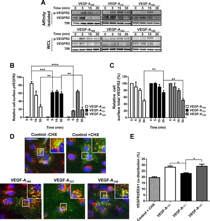
Fig. 2.
VEGF-A isoforms promote differential ligand-stimulated VEGFR2 internalization. (A) Endothelial cells were stimulated with either VEGF-A165, VEGF-A121 or VEGF-A145 (1.25 nM) for 5, 15 or 30 min before cell surface biotinylation, affinity isolation and immunoblot analysis of whole cell lysates (WCL) or biotinylated cell surface proteins (Affinity isolated). (B,C) Quantification of cell surface (B) activated VEGFR2-pY1175 or (C) mature total VEGFR2 levels upon VEGF-A isoform stimulation. Transferrin receptor (TfR) was used as a loading control. (D) Endothelial cells were pre-treated with cycloheximide (CHX; 2 μg/ml) for 2 h prior to VEGF-A isoform stimulation (1.25 nM) for 30 min. Endothelial cells were fixed and processed for immunofluorescence microscopy; VEGFR2 (green), EEA1 (red), nuclei (blue). Scale bar, 20 mm. (E) Quantification of VEGFR2/EEA1 co-distribution upon VEGF-A stimulation. Error bars indicate ±s.e.m. (n≥3). *P<0.05, **P<0.01, ***P<0.001, ****P<0.0001.

