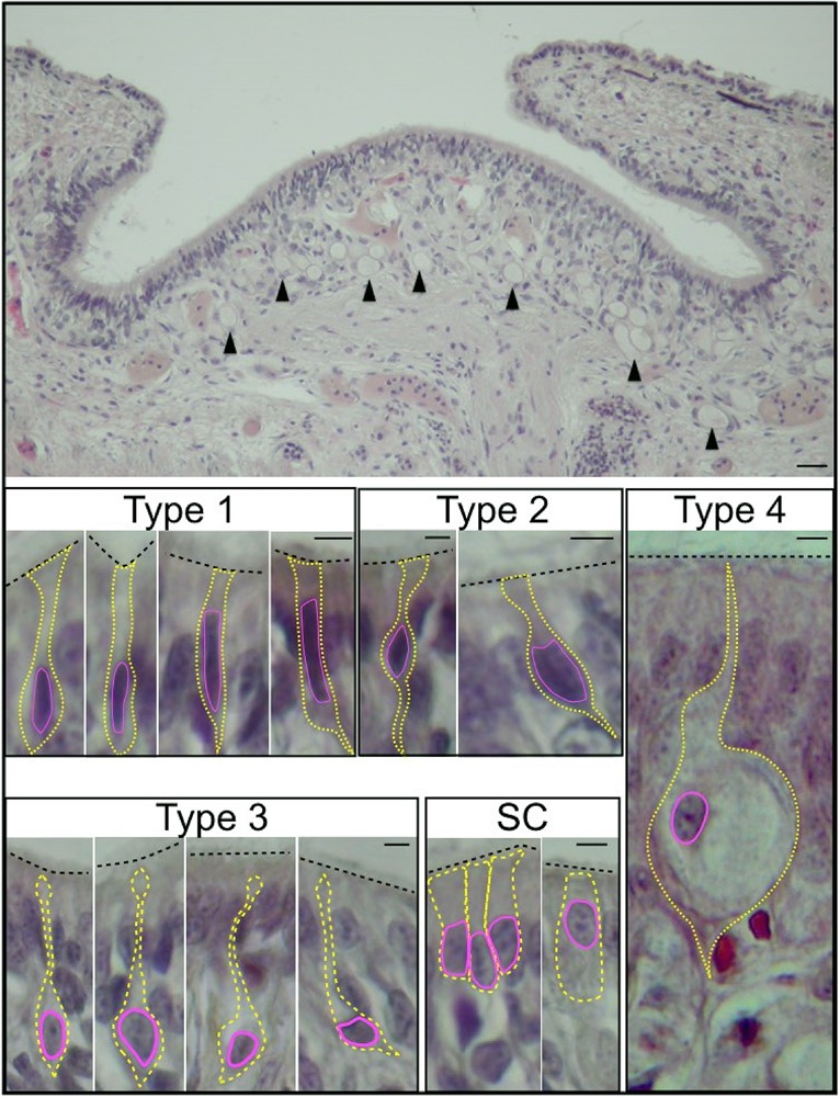
Fig. 2.

Transverse section of O. vulgaris olfactory organ. Top: the olfactory epithelium (scale bar=100 µm), arrowheads indicate vacuolated cells (type 4). The sensory cells are shown below: type 1 sensory cells have an elongated nucleus and minimal cytoplasm; type 2 sensory cells have a central nucleus and project to the epithelial surface and to the basal lamina; type 3 sensory cells have a soma occupied by a large nucleus and a long process directed to the surface; type 4 sensory cells are pear shaped cells with a large vacuole, an eccentric nucleus, minimal cytoplasm and a long projection which appears to terminate in cilia. Cylindrical sustentacular cells (SC) emerge onto the epithelial surface with an apical brush border of microvilli. Scale bars=5 µm.
