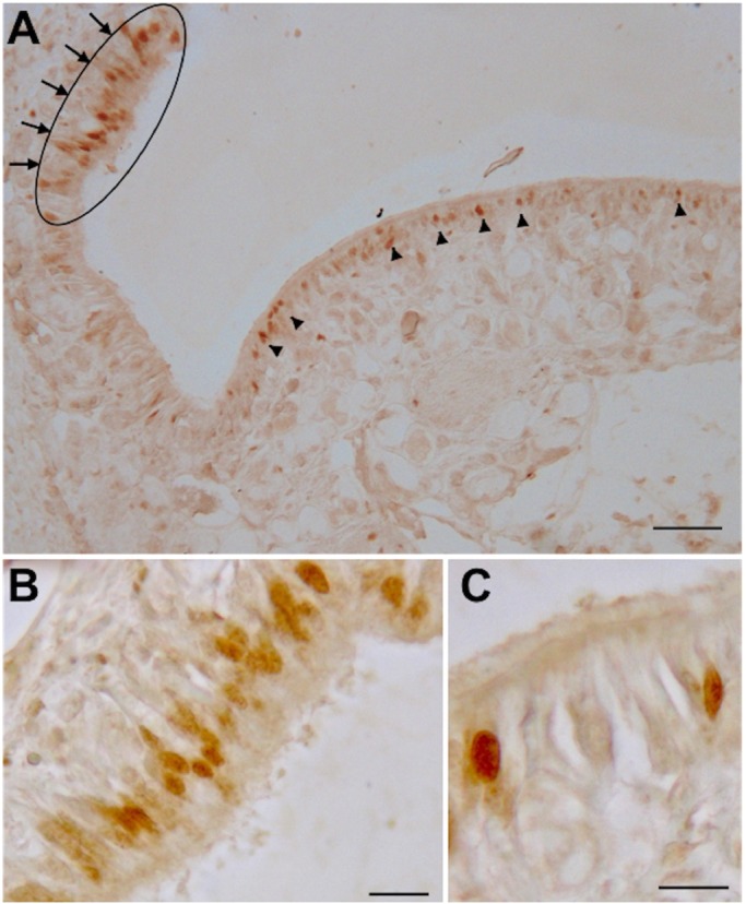
Fig. 4.

PCNA immunoreactivity on a transverse section of O. vulgaris olfactory organ. (A) Overview of the olfactory epithelium with several olfactory sensory neurons labeled. The arrowed oval indicates the most proliferative area with a concentration of PCNA immunoreactivity nuclei on the peripheral fold of the epithelium, the arrowheads indicate some scattered PCNA immunoreactivity nuclei on the central epithelium area. (B,C) Magnifications with PCNA immunoreactivity cells in the fold and into the olfactory protuberance, respectively, of the olfactory epithelia. Scale bars=100 µm in A, 10 µm in B,C.
