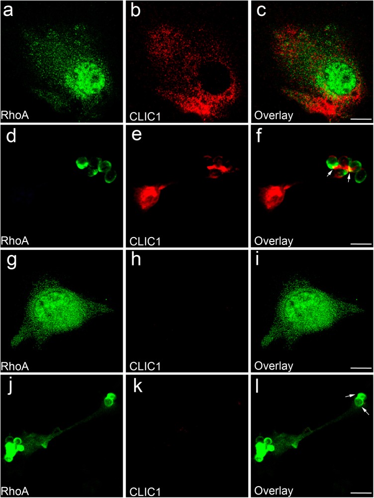
Fig. 1.
Phagocytosis triggers CLIC1 translocation to BMDC phagosomal membrane. (A-F) Immunofluorescence confocal microscopic images of resting CLIC1+/+ BMDCs (A-C) or BMDCs phagocytosing IgG-opsonised zymosan particles (D-F), stained with antibodies to RhoA (green) or CLIC1 (red). (G-L) Images of resting CLIC1−/− BMDCs (G-I) or BMDCs phagocytosing IgG-opsonised zymosan particles (J-L), stained with antibodies to RhoA (green) and CLIC1 (red). Both CLIC1 and RhoA appear on the phagosomal membrane after 5 min of phagocytosis in CLIC1+/+ BMDCs (F, arrows). Only RhoA can be identified on the phagosomal membrane of CLIC1−/− BMDCs (L, arrows). Scale bar: 10 µm.

