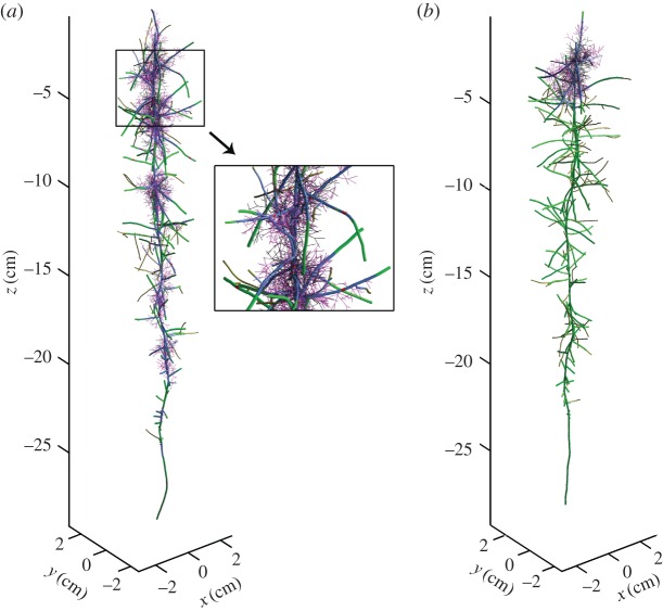
Figure 3.
Twenty-one day old simulated mycorrhizal root system with unconfined root growth and (a) dispersed inoculum with infection probability 0.15; close-up of part of the mycorrhizal root (b) concentrated inoculum with infection probability 1. Green, uninfected root segment; red, root segment infected by primary infection; blue, root segment infected by secondary infection; magenta, external hyphae up to 10 days old; black, dead roots or external hyphae.

