
Figure 3.
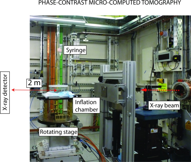
Experimental protocol for PC μCT. The inflation chamber (shown in Fig. 2) was immersed in a PBS bath, and the entire apparatus was placed on a rotating stage. An x-ray beam, produced by synchrotron radiation, was directed toward the ONH region. The x-ray detector was placed 2 meters behind the specimen. For PC μCT, a few millimeters of optic nerve were left on the eye.