
Fig. 7.
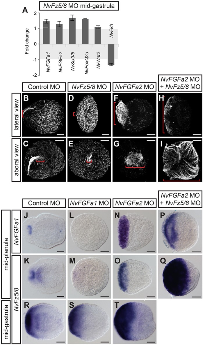
NvFz5/8 regulates the size of the apical organ by controlling FGF activity in the aboral-most domain. (A) RT-qPCR of NvFz5/8 MO-injected embryos at mid-gastrula (26 hpf). Fold changes of the relative expression levels of the indicated genes are shown; values between −1 and +1 mean no change (highlighted in light gray). Error bars represent the s.d. of three biological replicates. (B-I) Lateral views (aboral pole to the left; B,D,F,H) and aboral views (C,E,G,I) of the apical ciliary tuft visualized by anti-acetylated tubulin antibody staining of mid-planulae. The injected MOs are indicated above. The red brackets highlight the size of the apical tuft in the different conditions. (J-T) Lateral views (aboral pole to the left) of NvFGFa1 and NvFz5/8 in situ expression patterns in mid-planula (J-Q) and mid-gastrula (R-T) MO-injected embryos. The injected MOs and probes used are indicated above and on the left, respectively. Scale bars: 50 µm.