
Fig. 2.
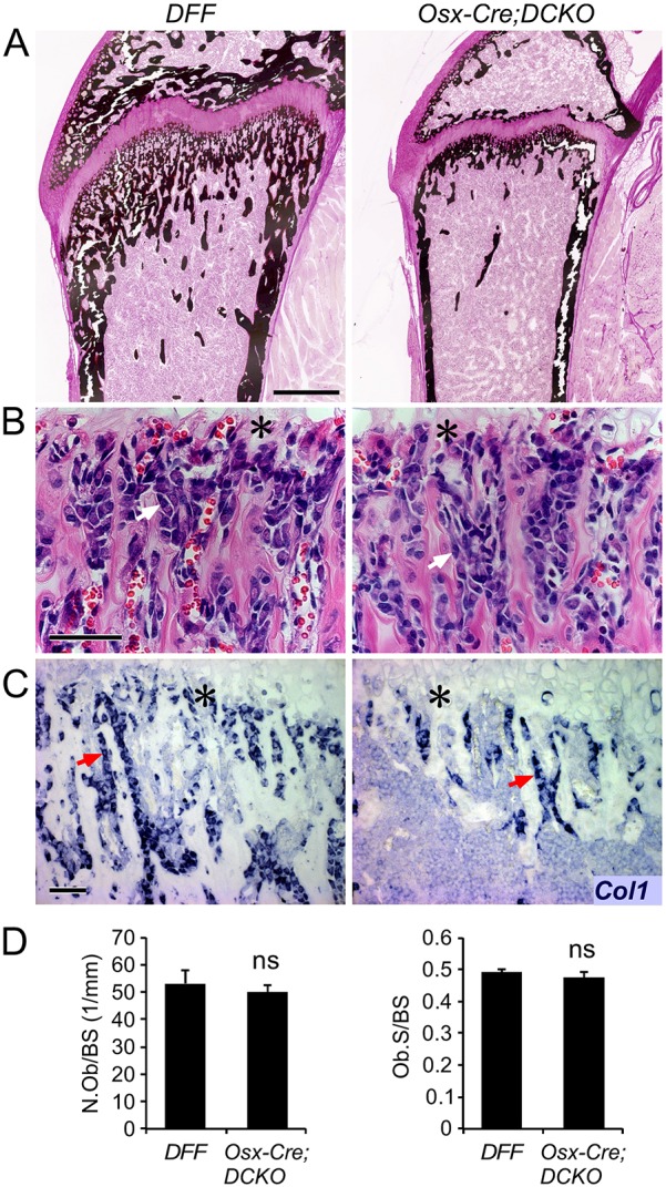
Decreased cortical and trabecular bone formation in Osx-Cre;DCKO mice. (A) Histology of the proximal tibia at P21, showing reduced mineralized bone (von Kossa stain) in Osx-Cre;DCKO mice. (B) Histology (H&E staining) showing normal osteoblast morphology in the trabecular region adjacent to the chondro-osseous junction (asterisk) in Osx-Cre;DCKO mice. (C) Type I collagen (Col1) expression detected by in situ hybridization in DFF and Osx-Cre;DCKO mice. (D) Histomorphometry of DFF and Osx-Cre;DCKO mice (n=3) showing normal osteoblast number per bone surface (BS) area and normal osteoblast surface per bone surface. Arrows (B,C) indicate osteoblasts. ns, non significant. Scale bars: A, 500 µm; B,C, 50 µm.